
Saturday, 29 November 2014
Friday, 28 November 2014

Tuesday, 25 November 2014
Digestive Lab
Label


Oil Test After


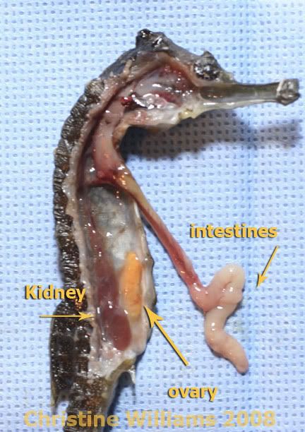
Stomach Vary in the Animal Kingdom
Everybody knows that humans have only one stomach which is apart of the digestive system however, it is not the same for certain animals. For example, cows, giraffes, deer, and countless more animals have stomachs with multiple compartments as a result they have four chambered stomachs which help them with their digestion. While other animals such as seahorses have a different digestive root, the food goes straight from their esophagus to their intestines. I choose this topic because until now I thought every animal had one stomach. After reading this article, I wonder how animals similar to seahorses break down their food?
 \
\
\Monday, 17 November 2014
Protein Lab
1. A peptide bond is found in the primary structure between the amino acids.
2. The secondary structure forms because of polar peptide bonds between the amino acids.
3. The interaction between the different R groups which are basically amino acids create the tertiary structure.
4. Two or more polypeptides which are stuck together create the quaternary structure. My protein shape looked differently than everybody else's proteins because everybody had different R groups therefore the interaction between their amino acids will cause the shapes to be different.
5. My protein would not have the same shape if I changed the primary structure because the interaction of the R groups would be different.
6. The highlighted parts represent the different R groups and this picture shows that the amino acid structure is not as straightforward as shown on our notes
This is the picture of my protein at the final stage.
Friday, 14 November 2014
Colon Cancer Surgery
After seeing countless pictures of the Colon Cancer surgery and numerous tweets which were tweeted during the operation, I was shocked to discover that the surgeons inserted a port which pumped carbon dioxide into the abdomen to create space because as well as a better view. In my opinion the most amazing part of the surgery was when the team put laparascope (tiny videos) into the abdomen to create videos which helped learn more about the abdomen from the inside. Some the parts that I learnt in class and I saw during the surgery was the colon. The most interesting part was when I found out one of the tools used in this surgery which was called grasper, it was interesting because it allowed the surgeons to move as well as grasp the colon without
puncturing it. Something new I learnt today that I did not know before was that the surgeons tattooed the tumor with ink to make it easier to find it.


puncturing it. Something new I learnt today that I did not know before was that the surgeons tattooed the tumor with ink to make it easier to find it.
Tuesday, 11 November 2014
Respiration Lab

This is a picture of two test tubes full of limewater. A student will use a straw and breath into the left test tube while another student will areate the limewater in the right test tube.

The test tube which was areated did not change color because it stayed neutral however, there were some bubbles. While the other test was not transpearnt anymore instead, the color became white as a result of contact with carbon dioxide from the student.

Both small beakers are full of 25ml of distilled water however, one beaker will be areated using a pipette while a student will breathe into the water in the other beaker for five minutes. After those five minutes one must add one drop of universal indactor to both beakers.

The beaker on the left came in contact with carbon dioxide and it changed color from white to yellow due to the universal indicator however, this proves that it has become more acidic in PH level. While, the beaker on right was areated and it changed color from white to green which proves that it is still neutral
Friday, 7 November 2014
Ankle Roll
An ankle roll can occur to anybody at anytime in the world, this injury does not have to happen during physical activities such as basketball, you could roll your ankle if you step on an uneven surface or step on an angle. Ankle joints as wells as the bone are held together in place by ligaments which is an elastic structure like an rubber band that protects the ankle joints from twisting or rolling. When a person rolls their ankle, their ligaments are stretched beyond their normal position as a result tearing the elastic fibers. Also, when a person rolls their ankle and if the force was too great then they can completely tear their ligaments which may acquire the person to undergo surgery. Some people believe they have rolled their ankles so they do not go to the doctor however there is always a chance that you could have a broken bone in the ankle or foot. A broken bone can have similar symptoms of pain and swelling. I choose this topic because I have a history of rolling ankles.

Severity | Physical Examination Findings | Impairment | Pathophysiology | Typical Treatment* |
|---|---|---|---|---|
Grade 1
|
Minimal tenderness and swelling
|
Minimal
|
Microscopic tearing of collagen fibers
|
Weight bearing as tolerated
No splinting/casting
Isometric exercises
Full range-of-motion and stretching/ strengthening exercises as tolerated
|
Grade 2
|
Moderated tenderness and swelling
Decreased range of motion
Possible instability
|
Moderated
|
Complete tears of some but not all collagen fibers in the ligament
|
Immobilization with air splint
Physical therapy with range-of-motion and stretching/ strengthening exercises
|
Grade 3
|
Significant swelling and tenderness
Instability
|
Severe
|
Complete tear/ rupture of ligament
|
Immobilization
Physical therapy similar to that for grade 2 sprains but over a longer period
Possible surgical reconstruction
|
Tuesday, 4 November 2014

Subscribe to:
Comments (Atom)